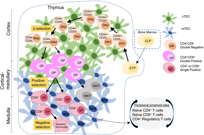
Figure 1: The development of T cells in the thymus.
Bone marrow (BM)-derived lymphoid progenitor cells enter the thymus to begin commitment to the T cell lineage, becoming double negative ‘DN’ thymocytes (tan-orange) based on the lack of expression of CD4 and CD8 co-receptors. DN thymocytes progress through sequential DN1-4 stages, as defined by the coordinate expression of CD44 and CD25 on the cell surface. The T cell receptor (TCR) β-chain is expressed at DN3, triggering progression and maturation to double positive ‘DP’ thymocytes (pink) expressing both CD4 and CD8 co-receptors. Positive selection delineates selection of thymocytes into the CD4, T-helper, or CD8, cytotoxic T cell lineage to become single positive ‘SP’ CD4 or CD8 T cells (maroon). After positive selection, SP CD4+ or CD8+ T cells migrate to the medulla to go through negative selection mediated by mTECs, where autoreactive SP T cells are deleted by apoptosis while SP T cells that pass negative selection are exported to the periphery. This process of thymopoiesis results in population of peripheral blood and lymphoid sites with naive CD4+ and CD8+T cells and CD4+ regulatory T cells (Tregs).

