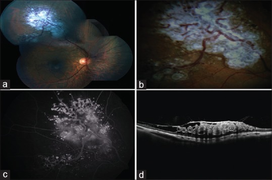
Figure 1.

(a and b) Color fundus photograph of the right eye showing saccular blood vessels with surface gliosis; appearing like a “cluster of grapes”. (c) Fluorescein angiography of the right eye showing delayed hyperfluorescence in the aneurysms with plasma–erythrocyte separation; termed as fluorescein caps. (d) Optical coherence tomography shows lobulated spaces in inner retina correspond to the aneurysms and overlying epiretinal membranes with traction
