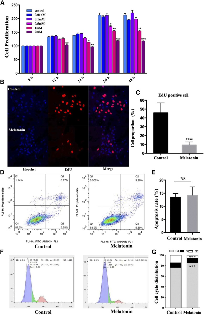
Fig. 1.
Effect of melatonin on cell proliferation in porcine intramuscular preadipocytes. A: The cells were treated with various melatonin concentrations (0, 0.01, 0.1, 0.5, 1, and 2 mM) at various times 0, 12, 24, 36, or 48 h. After treatment, cell proliferation was estimated by Cell Counting Kit-8 assay. Results are shown as a relative percentage of untreated cells at 0 h (n = 4 per group). B: Detection of intramuscular preadipocyte cellular activity by EdU staining after treatment with or without 1 mM of melatonin for 24 h. The proliferating nuclei were stained red with EdU, while the nuclei of all cells were stained blue with Hoechst for 30 min (scale bar = 100 μm). C: Quantification of the proportion of EdU-positive cells. D: The effect of melatonin on the cell cycles and apoptosis of preadipocytes. Cells were stained with PI and annexin V, and then the apoptosis rate was evaluated by flow cytometry and displayed as column charts after quantification (E). F: Cell cycle analysis. Cells were stained with PI, and then detected by flow cytometry and displayed as column charts after quantification (G). Data are represented as the mean ± SEM (*P < 0.05; **P < 0.01; ***P < 0.001; n ≥ 3).

