Fig. 6.
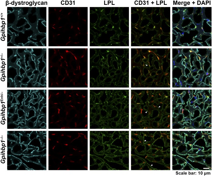
LPL is partially mislocalized in the heart in Gpihbp1Enh/− mice, with increased amounts of LPL in the interstitial spaces near the surface of cardiomyocytes. Confocal microscopy studies were performed on sections of heart stained with antibodies for β-dystroglycan (cyan), CD31 (red), and LPL (green). β-Dystroglycan is located along the surface of cardiomyocytes. In comparing confocal images from Gpihbp1+/+ and Gpihbp1Enh/− mice, we observed more LPL outside of capillaries in Gpihbp1Enh/− mice (colocalizing with β-dystroglycan) (arrowheads in the CD31/LPL merged image point to several such regions). An even greater amount of interstitial LPL (colocalizing with β-dystroglycan) was observed in sections from Gpihbp1−/− mice (arrowheads). A small amount of LPL was mislocalized to the interstitial spaces in Gpihbp1+/− mice (arrowhead). DNA was stained with DAPI (blue). Scale bar, 10 μm.

