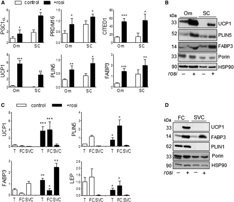
Fig. 1.
Rosi induced brite phenotypes in both Om adipose tissues and subcutaneous human adipose tissues. A, B: mRNA and protein levels of genes known to be more abundantly expressed in brown and brite than white adipocytes were measured in Om adipose tissues and abdominal subcutaneous (SC) human adipose tissues that had been cultured without or with Rosi (1 μM) for 7 days. C, D: After treating Om adipose tissue without or with Rosi, fat cells (FC) and stromal vascular cells (SVC) were isolated with collagenase digestion and mRNA and protein levels were measured in tissue (T), FCs, and SVCs. Data are expressed as the mean ± SEM of five to nine experiments [BMI, 37.5 ± 3.7 kg/m2 (range 29–54 kg/m2); age, 50.8 ± 4.0 years (range 36–71 years); seven female/two male]. *P < 0.05, **P < 0.01, ***P < 0.001 (paired t-tests after one- or two-way ANOVA).

