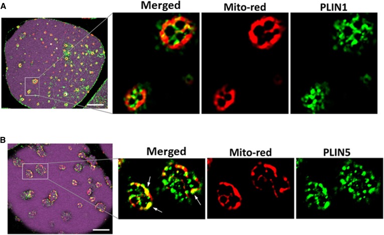
Fig. 6.
PLIN5 coated the small droplets and colocalized with mitochondria in brite adipocytes. Adipocytes isolated from Om tissues after 7 day culture without or with Rosi were stained with LipidTOX-Deep Red and Mitotracker-Red and used for immunostaining of PLIN1 (A) and PLIN5 (B), as described in the Materials and Methods. White arrows indicate where PLIN5 colocalized with mitochondria. White scale bars = 10 μm.

