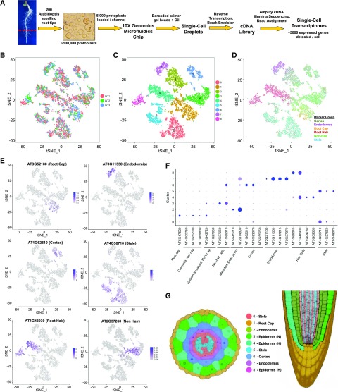
Figure 1.
Isolation and cluster analysis of single-cell transcriptomes from wild-type Arabidopsis roots. A, Workflow used for scRNA-seq to obtain transcriptomes from individual Arabidopsis root cells. B, tSNE projection plot showing dimensional reduction of the distribution of 7522 individual wild-type cell transcriptomes from three biological replicates. Cell transcriptomes derived from each replicate are indicated by different colors (red = replicate 1; green = replicate 2; blue = replicate 3). C, tSNE projection plot showing nine major clusters of the 7522 individual wild-type root cell transcriptomes. D, tSNE projection plot showing the combined transcript accumulation from all marker genes tested (listed in Supplemental Table S3) on the 7522 wild-type transcriptomes, organized by the tissue/cell type of the marker gene group (cortex, endodermis, root cap, root-hair, nonhair, and stele marker gene sets). E, tSNE projection plots showing transcript accumulation for specific tissue/cell marker genes in individual cells. Color intensity indicates the relative transcript level for the indicated gene in each cell. The tissue/cell types known to preferentially express each marker gene are given in parentheses (details available in Supplemental Table S3). Additional marker gene plots are provided in Supplemental Figure S2. F, Accumulation of marker gene transcripts by cluster. This dot plot indicates the fraction of cells in each cluster expressing a given marker (dot size) and the level of marker gene expression (dot intensity) for 24 genes known to exhibit preferential expression in distinct tissue/cell types (Supplemental Table S3). G, Assignment of cell clusters to root tissues. Depictions of transverse (left) and longitudinal (right) sections of the Arabidopsis primary root showing tissues in colors corresponding to the nine-cluster plot in C. N, Nonhair cells; H, root-hair cells. Note that cells corresponding to cluster 8 are not shown on the root images, because this cluster was found to contain mature root-hair cells which are not represented in these root images.

