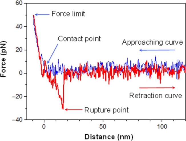
Figure 3.

When approaching the cantilever tip, (blue curve), the distance between the tip and the cell surface decreases. At the contact point, the tip touches the cell membrane. After the contact (left side of the contact point), the tip gently presses on the cell membrane. When the force limit (which is about 50 pN here) is reached, the tip is retracted from the cell surface. If the antibody binds to the heparan sulfate, the cantilever tip is pulled downwards (red curve) until the 2 molecules are separated at a critical force. After the rupture, the cantilever recovers its resting state. If there is no binding between antibody and heparan sulfate during the contact, the retraction curve looks similar to the approaching curve [Colour figure can be viewed at wileyonlinelibrary.com]
