
Figure 6.
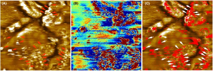
Heparan sulfate mapping of the stripped abdominal skin sample using a cantilever tip functionalized with the anti‐heparan sulfate antibody. The scan size is 1 μm × 1 μm. (A) Topography image. The height scale is 12 nm for the full color range. (B) Simultaneously acquired recognition map. Blue to orange reflects no recognition and dark red corresponds to a recognition event. (C) An overlay of the topography and the recognition map showed that heparan sulfate is located on surface protrusions (bead‐like structures, indicated with arrows) [Colour figure can be viewed at wileyonlinelibrary.com]