Figure 8.
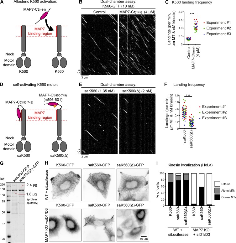
MAP7-Ct can activate kinesin-1. (A and D) Overview of kinesin-1 and MAP7 constructs used for experiments. (B and E) Kymographs of a dual-chamber in vitro experiment, where equal concentrations of K560-GFP motors were added to chambers with or without MAP7-Ct(mini) (B), or with the indicated concentrations of saK560/saK560(Δ) (E) on dynamic MTs. (C and F) Landing frequencies quantified per MT and corrected for MT length, time of acquisition, and kinesin concentration. Each independent dual-chamber experiment is color-coded; n = 32 and 29 MTs (C) and n = 73 and 69 MTs (F), all from three independent experiments. ***, P < 0.001, Mann–Whitney U test. (G) Analysis of purified saK560 proteins by SDS-PAGE. Protein concentrations were determined from a single gel using BSA standard. (H) Widefield images of overexpressed GFP-tagged kinesin constructs in control or MAP7 KO + siMAP7D1/D3 HeLa cells. (I) Quantification of kinesin localization categorized as diffuse, along MTs, or at corner MTs. n = 203, 172, 198, 241, 186, and 234 cells from three independent experiments.

