Figure 5.
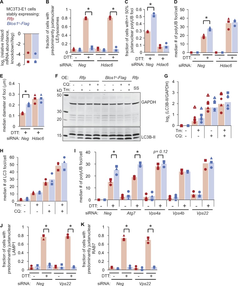
Protein aggregate clearance during ER stress relies on HDAC6 and the ESCRTs. (A–E) We transfected cells with siRNAs targeting Neg or Hdac6, and then treated with DTT (2 mM, 4 h). We analyzed Hdac6 mRNA levels by qPCR (A), LE/lysosome repositioning by LAMP1 immunostaining (B), or polyubiquitin foci (C–E). (F) We treated cells with Tm (6 µg/ml) or serum-free media as a control (18 h). We added CQ (120 µM), where indicated for the final 2 h, and then analyzed LC3B processing by immunoblot. (G) Quantification of five independent experiments as in F. (H) We treated cells as in F and G, fixed and stained with an LC3B antibody, and counted LC3B foci. (I) We transfected cells with siRNAs targeting autophagy components or control siRNAs (Neg), then treated with DTT (2 mM, 4 h) and analyzed polyubiquitin foci. RNAi controls are shown in Fig. S3. (J and K) We depleted cells of Vps22, treated with DTT (2 mM, 4 h), and analyzed LE/lysosome repositioning by either LAMP1 (J) or RAB7 (K) immunostaining. All panels: *P < 0.05 for Rfp versus Blos1s cells, using paired t tests with corrections for multiple comparisons; P values between 0.05 and 0.15 are shown. n = 3 except for G. OE, overexpressed; SS, serum starvation.

