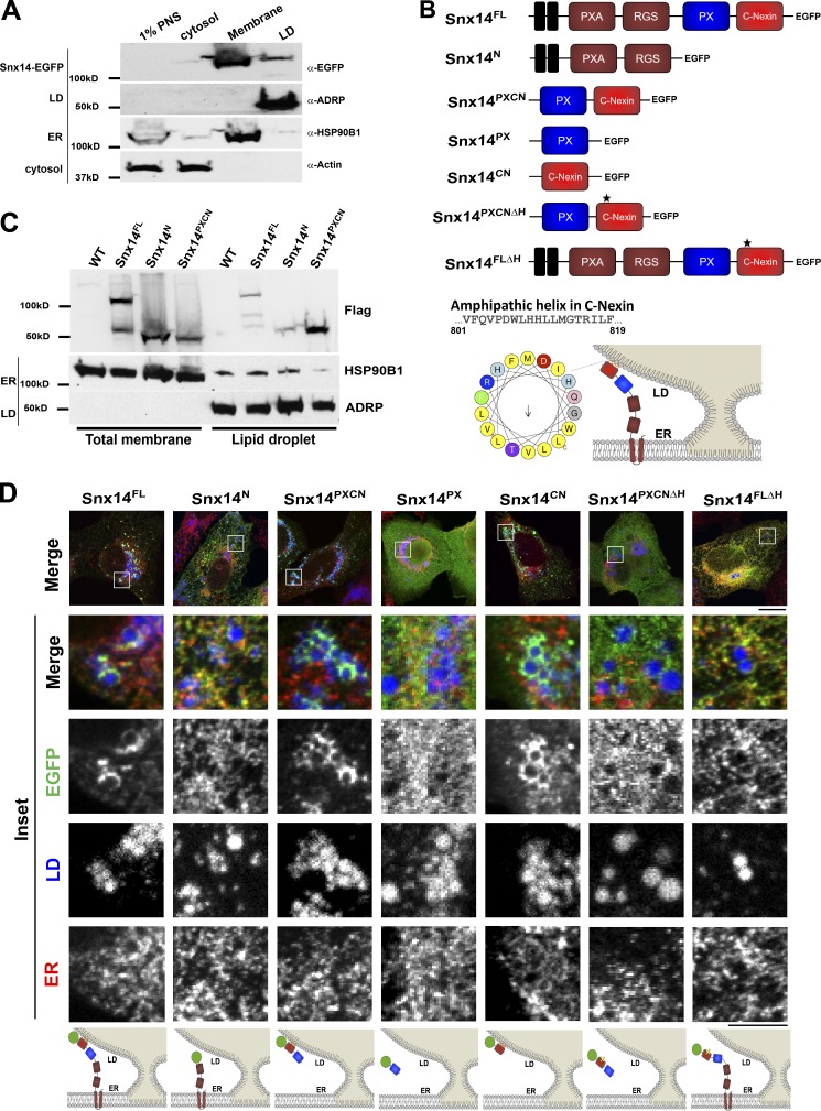
Figure 2.
Snx14 is topologically anchored in the ER and interacts with LDs in trans. (A) LD flotation assay of OA-treated U2OS cells expressing Snx14EGFP. Lanes indicate postnuclear supernatant (PNS), cytosol, total membrane, and LD float fractions. (B) Schematic diagram of Snx14 fragment constructs tagged with EGFP. Snx14FL depicts the full-length human Snx14. Snx14N is the N-terminal fragment from the start that includes PXA and RGS domains. Snx14PXCN includes the PX domain and C-Nexin domains. Snx14PX consists of PX and Snx14CN represents C-Nexin domain. An AH in the C-Nexin domain is identified as depicted in the schematic diagram. Snx14PXCNΔH indicates the PX and C-Nexin domain from which the AH is deleted. Snx14FLΔH depicts the full-length Snx14 with AH deletion. (C) Western blot showing distribution of Snx14FL, Snx14N, and Snx14PXCN tagged with 3XFlag among total membrane and LD fractions following OA treatment. (D) U2OS cells transfected with Snx14FL, Snx14N, Snx14PXCN, Snx14PX, Snx14CN, Snx14PXCNΔH, and Snx14FLΔH, respectively, and treated with OA for 16 h. CoIF staining with α−EGFP (green) and α-HSP90B1 (ER marker, red) and LDs stained with MDH (blue) and imaged by confocal microscopy. Scale bar = 20 µm. Inset scale bar = 5 µm. Cartoons represent the localization of the respective Snx14 fragments with respect to ER and LD.

