Figure 8.
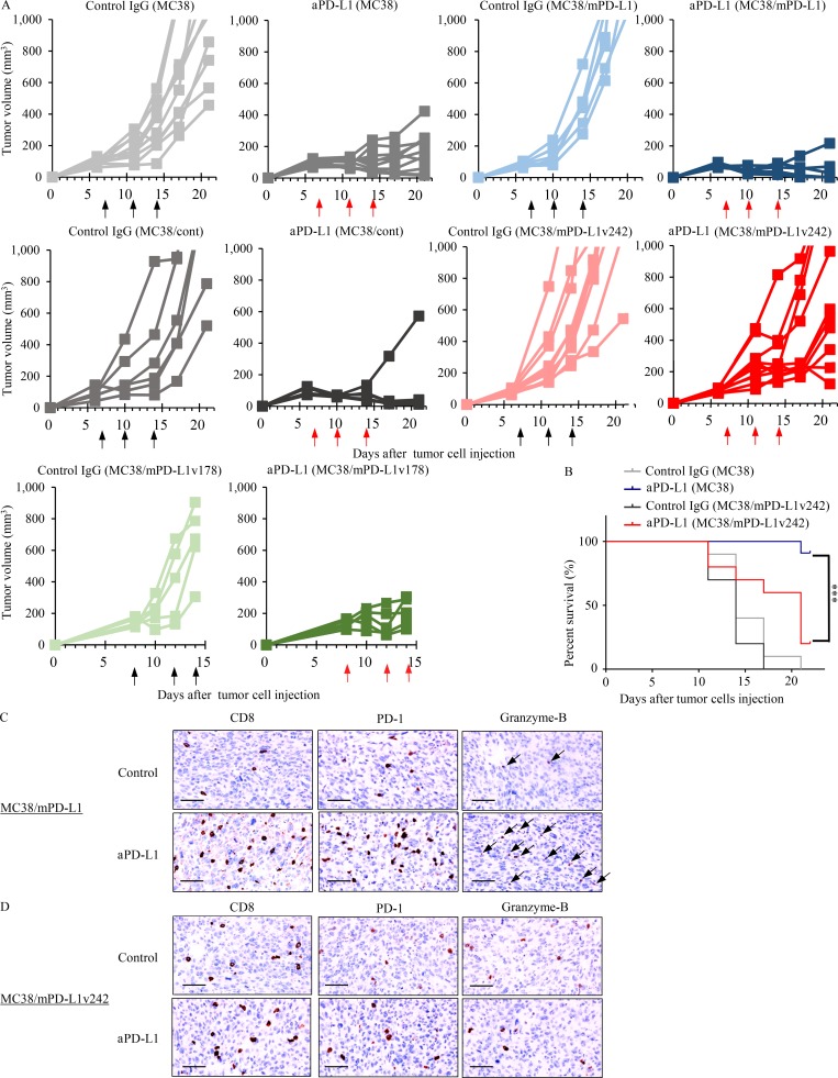
sPD-L1 splicing variants mediate the resistance to PD-L1 blockade in MC38 syngeneic mouse model. (A) C57BL/6 mice bearing MC38/cont (n = 6), MC38/mPD-L1 (n = 6), MC38 (n = 10), MC38/mPD-L1v242 (n = 10), and MC38/mPD-L1v178 (n = 5) were intraperitoneally administrated 50 µg/mouse control IgG or 35 µg/mouse aPD-L1 antibody. The schedules for treatment are indicated by black arrows (control IgG) and red arrows (aPD-L1). Tumor volume is plotted individually. (B) Kaplan–Meier survival curves for mice bearing MC38 or MC38/mPD-L1v242. The survival curves were compared by applying the Gehan–Breslow–Wilcoxon test. ***, P < 0.001. (C and D) Representative IHC staining of mouse CD8, PD-1, and granzyme B was performed on day 21 for MC38/mPD-L1 (C) and MC38/mPD-L1v242 (D) xenograft tumors. Bars, 20 µm. Each experiment was independently performed twice, yielding similar results.

