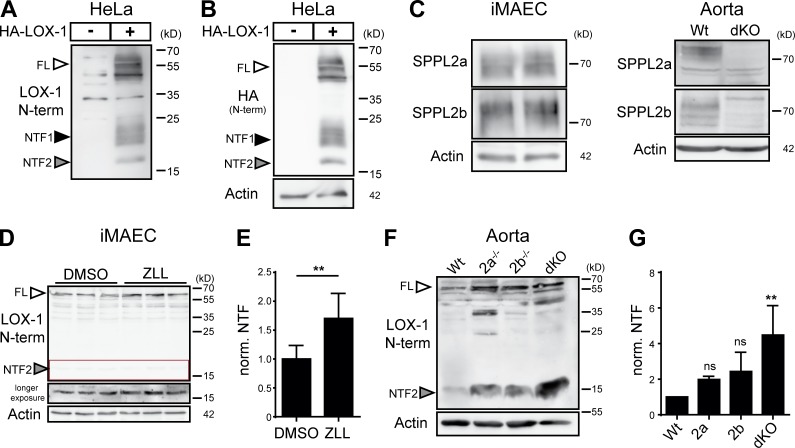
Figure 4.
Endogenous LOX-1 is processed by SPPL2a and SPPL2b. (A and B) Evaluation of functionality of the newly generated antibody against the N terminus of murine LOX-1 in transfected HeLa cells. (A) Blot probed with new antibody. (B) Same blot as in A probed with anti-HA. (C) Expression of SPPL2a and SPPL2b was demonstrated in iMAECs and aortic lysates by Western blotting. (D) iMAECs were treated for 24 h with either 40 µM ZLL or DMSO before Western blot analysis. (E) Quantification of D. N = 2, n = 6. Student’s t test. (F) LOX-1 NTF levels were analyzed in aortic lysates from WT, SPPL2a-, SPPL2b-, or dKO mice by Western blotting. (G) Quantification of F. N = 4, n = 4. One-way ANOVA with Tukey’s post hoc testing. **, P ≤ 0.01; ns, not significant. N, the number of independent experiments; n, the number of individual samples for quantification. All data are shown as mean ± SD.

