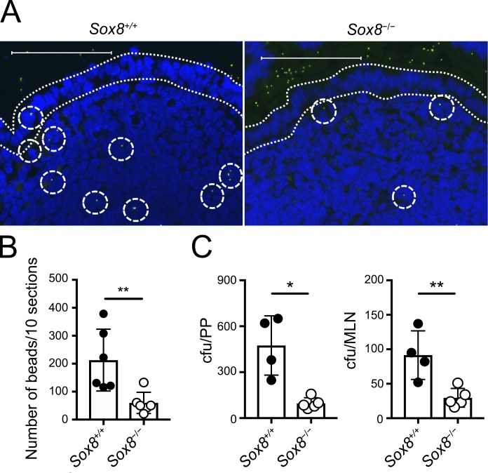
Figure 4.
The loss of Sox8 causes decreased uptake of luminal nanoparticles into the follicle. (A and B) Green fluorescent latex beads (200-nm diameter) were orally administered to Sox8+/+ or Sox8−/− mice. 3 h later, two PPs were collected from the jejunum. Cryosections of PPs were examined by fluorescence microscopy, and the number of particles was counted manually. (A) Representative images of cryosections. Dotted lines indicate FAE. Dotted circles indicate fluorescent particles. Bars, 100 µm. (B) Quantification of particles in PPs. **, P < 0.01; Student’s t test; n = 6 per genotype. (C) Mice were orally infected with 5 × 10⁸ CFU of S. Typhimurium (ΔaroA); 24 h later, PPs and MLNs were collected and tissue homogenates were prepared. The colonies of culturable bacteria in the tissue homogenates were counted and are shown as colony-forming units. **, P < 0.01; *, P < 0.05; unpaired two-tailed Student’s t test; n = 4 from Sox8+/+ mice, n = 5 from Sox8−/− mice. Data are representative of two independent experiments. All values are presented as the mean ± SD.

