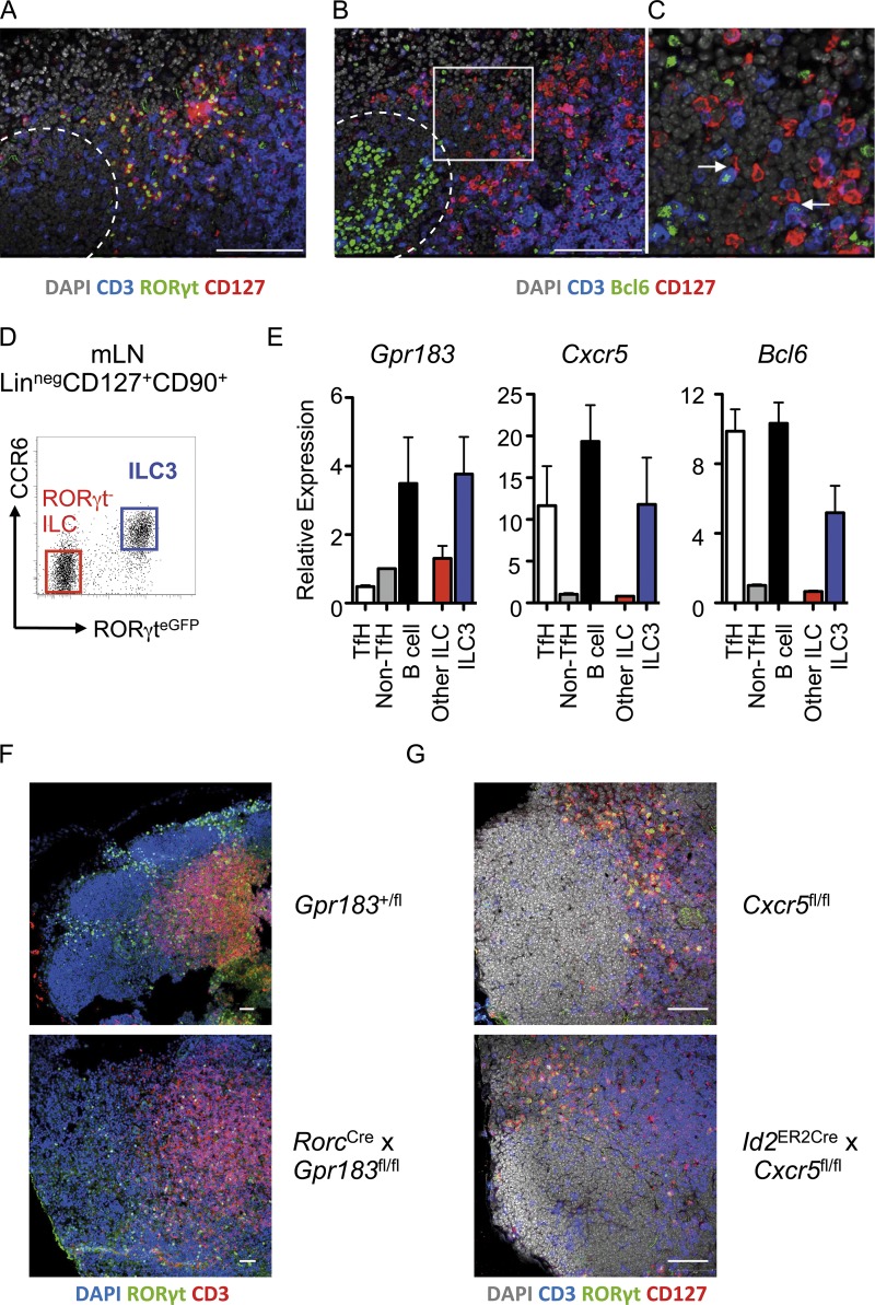
Figure 1.
LTi-like ILC3 localize to interfollicular regions of the mLN and interact with TfH cells. (A and B) Serial sections of mLN demonstrating (A) positioning of RORγt+CD127+ CD3− ILC3 and (B) colocalization of CD127+CD3− ILC with CD127−CD3+Bcl6+ TfH cells in wild-type C57BL/6 mice (n = 6), representative of two independent experiments. Dotted line indicates follicular border. Bars, 100 µm. (C) Higher magnification image of insert indicated in B. Arrows indicate contacts between ILC3 and TfH cells in interfollicular region. (D and E) (Live CD45+/Linneg/CD127+) RORγteGFP+ LTi-like ILC3 (n = 3; blue) and RORγt-negative ILC (n = 3; red) were sorted from the mLN of RORγteGFP mice along with (CD3−CD4+) PD-1+ICOS+ TfH (n = 6), PD-1−ICOS− conventional T cells (n = 6), and CD19+B220+MHCII+ B cells (n = 6; D) and assessed for expression of Gpr183, Cxcr5, and Bcl6 (E), with data representative of two independent experiments. (F) Localization of DAPI+RORγt+ CD3− ILC3 in the mLN of Gpr183+/fl control mice or RorcCre × Gpr183fl/fl mice (n = 5 per group), representative of two independent experiments. (G) Localization of CD127+RORγt+CD3− ILC3 in the mLN of Cxcr5fl/fl control mice (n = 6) or Id2CreERT2 × Cxcr5fl/fl mice (n = 7) treated four times with tamoxifen and rested for 1 mo, representative of two independent experiments. Bar, 50 µm. All data shown as mean ± SEM.

