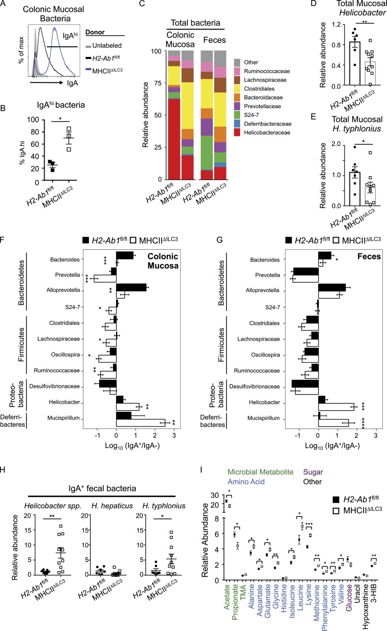
Figure 3.
Lack of ILC3 antigen-presentation results in dysregulated IgA coating of mucosal-dwelling bacteria in the colon. Bacteria isolated from the colonic mucosa of IgMi mice, which lack endogenous secreted IgA, were cocultured with fecal supernatants containing free IgA. (A and B) Representative histogram of IgA labeling of bacteria without incubation (unlabeled; gray fill) or cultured with fecal supernatant isolated from MHCIIΔILC3 mice (n = 3, blue line) and H2-Ab1fl/fl littermate controls (n = 3, black line; A) and frequencies of IgA-labeled bacteria (B), representative of two independent experiments. (C) Quantification of total bacterial families with a relative abundance >2% in the colonic mucosa or feces. (D and E) Relative abundance of Helicobacter spp. (D) or H. typhlonius (E) in total colonic mucosal bacteria preparations from C. (F and G) IgA-seq and log10 (IgA+/IgA−) enrichment score for major taxa with an abundance of at least 1% across all samples in the colonic mucosa (F) and feces (G). Statistical analysis was performed as detailed in the Materials and methods section and additionally confirmed via Student’s t test. (H) Relative abundance of total Helicobacter spp., H. hepaticus, and H. typhlonius in IgA+ fecal bacterial samples of MHCIIΔILC3 and H2-Ab1fl/fl mice. (C–H) Analysis of MHCIIΔILC3 (n = 10) and H2-Ab1fl/fl (n = 7) mice, with data pooled from two independent experiments. (I) Relative abundance of metabolites in the feces of MHCIIΔILC3 (n = 14) and H2-Ab1fl/fl (n = 9) mice, representative of pooled data from three independent experiments. Metabolites selected as significant via multivariate analysis and individual Student’s t test comparisons performed. All data shown as mean ± SEM; all statistical analyses performed by Student’s t test; *P ≤ 0.05; **P ≤ 0.01; ***P ≤ 0.001; ****P ≤ 0.0001.

