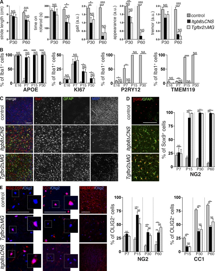
Figure 2.
Motor deficits and glial abnormalities in mice with deficient αVβ8 or TGFβ signaling to microglia. (A) Identical neuromotor symptoms in adult (P30–60) Itgb8ΔCNS and Tgfbr2ΔMG mice including waddling unstable gait with shortened stride length, reduced time on rotarod, unkempt appearance, and tremors. See also Videos 1 and 2. (B) Persistent expression of APOE and KI67 and reduced expression of P2RY12 and TMEM119 over time in microglia from Itgb8ΔCNS and Tgfbr2ΔMG mice. See accompanying Fig. S4. (C) Overlapping astrogliosis, microgliosis, and reduced MBP staining in P30 Itgb8ΔCNS and Tgfbr2ΔMG mice compared with controls. (D) Increased percentage of GFAP+SOX9+ astrocytes in Itgb8ΔCNS and Tgfbr2ΔMG mice over time; quantified on right. See accompanying Fig. S4. (E) Increased percentage of OLIG2+NG2-DSR+ OPC, and reduced mature OLIG2+CC1+ oligodendrocytes over time (quantified on right) and reduced staining for mature myelin marker TMEM10 in Itgb8ΔCNS and Tgfbr2ΔMG mice compared with controls. See accompanying Fig. S4. Bars, 100 µm. Error bars indicate SE. *P < 0.05; **P < 0.005; ***P < 0.0005; ****P < 0.0001. Student’s t test. n = 4 animals for all groups. Behavioral analysis: ANOVA with Tukey’s post hoc test; n = 6 animals for all groups. a.u., arbitrary units.

