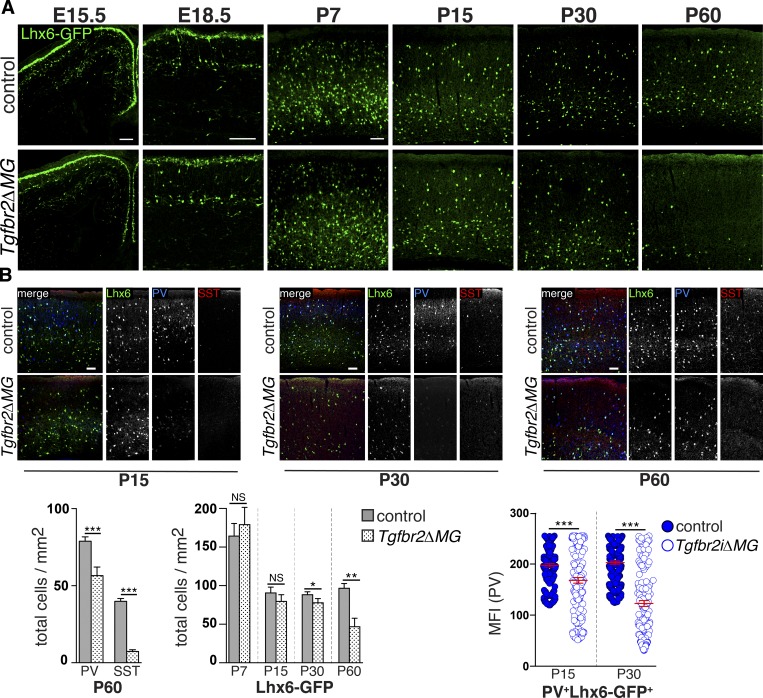
Figure 3.
Interneuron abnormalities in mice with deficient αVβ8 or TGFβ signaling to microglia. (A) Delayed loss of Lhx6-GFP+ cortical interneurons in Tgfbr2ΔMG; quantification below. (B) Reduced PV expression in Lhx6-GFP+ interneurons at P15 and P30, before the reduction in the numbers of these cells; quantification below. See also Fig. S4. Bars, 100 µm. Error bars indicate SE. *P < 0.05; **P < 0.005; ***P < 0.0005. Student’s t test. n = 4 animals for all groups. MFI, mean fluorescence intensity.

