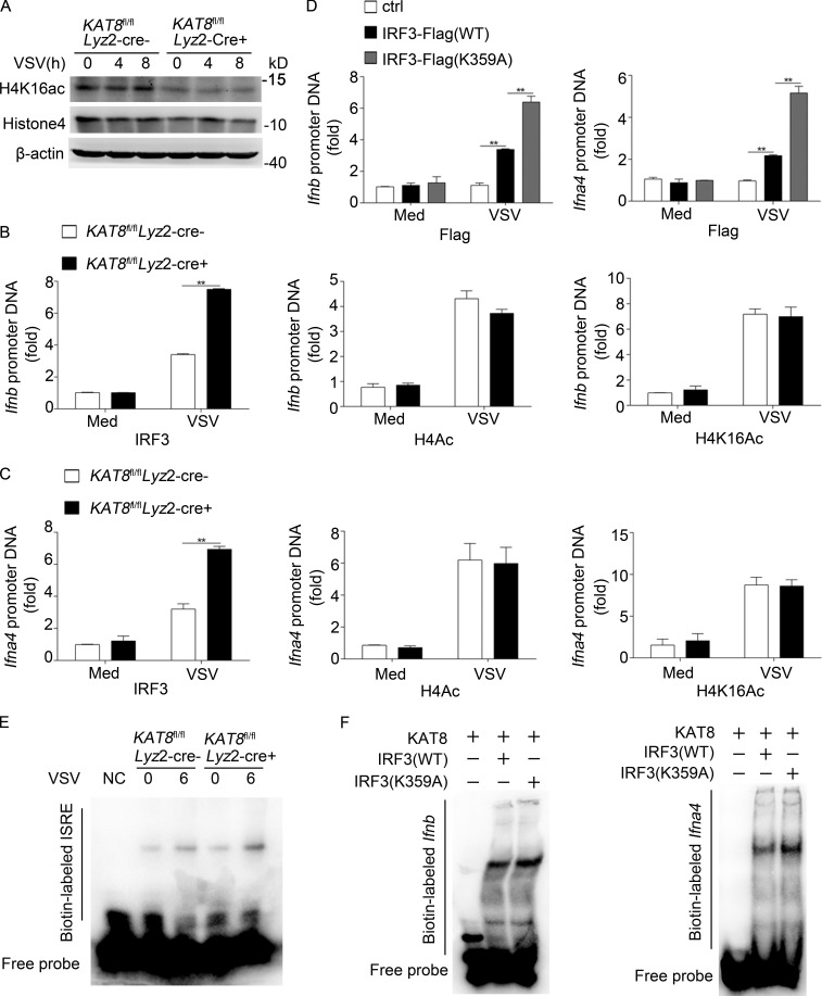
Figure 7.
KAT8 abrogates the abundance of IRF3 at IFN-I promoters. (A) Immunoblot analysis of H4K16ac, histone 4, or β-actin in KAT8fl/flLyz2-Cre− or KAT8fl/flLyz2-Cre+ peritoneal macrophages infected with VSV (1 MOI) for the indicated times. (B) ChIP analysis of IRF3, H4Ac, or H4K16Ac at the Ifnb promoter in KAT8fl/flLyz2-Cre− or KAT8fl/flLyz2-Cre+ peritoneal macrophages infected for 6 h with VSV (1 MOI) or left untreated. (C) ChIP analysis of IRF3, H4Ac, or H4K16Ac at Ifna4 promoter in KAT8fl/flLyz2-Cre− or KAT8fl/flLyz2-Cre+ peritoneal macrophages infected for 6 h with VSV (1 MOI) or left untreated. (D) ChIP analysis of IRF3 at Ifnb or Ifna4 promoter in MEF cells from IRF3-deficient mice overexpressed with WT or mutant IRF3 for 24 h and then infected with VSV (1 MOI) for 6 h. (E) EMSA of nuclear extract from KAT8fl/flLyz2-Cre− or KAT8fl/flLyz2-Cre+ peritoneal macrophages infected with VSV (1 MOI) for 6 h. The ISRE motif was biotin labeled. (F) Analysis of recombinant IRF3 WT or IRF3 mutant and IFN interaction in an EMSA assay. The Ifnb and Ifna motifs were biotin labeled. **, P < 0.01; two-tailed Student’s t test (B–D). Data are representative of three independent experiments with similar results (A, E, and F) or are from three independent experiments (B–D; mean ± SEM).

