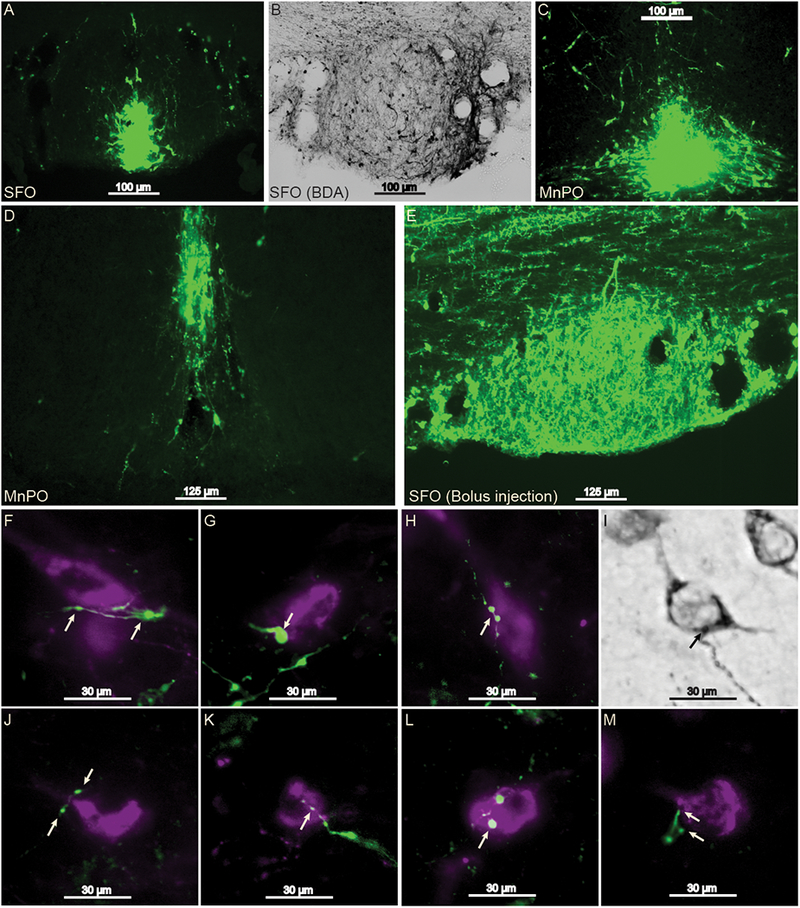
Figure 1 -.

Projections from the SFO and dorsal MnPO to Ox neuron cell bodies in the hypothalamus. 10× magnification of the focal point of iontophoresis injection sites in the SFO (panels A and B; one set of tissue was analyzed with DAB rather than immunofluorescence), dorsal MnPO (C and D), and a bolus injection in the SFO (E). All of the injection sites yielded anterograde labeling that consisted of varicosities or axon terminals in apposition with Ox cell bodies (panels F-M, images taken at 40× magnification). Arrows indicate sites of putative synaptic contact. Green represents BDA labeling and magenta represents Ox labeling. MnPO, median preoptic nucleus; Ox, orexin; SFO, subfornical organ.
