Figure 2 –

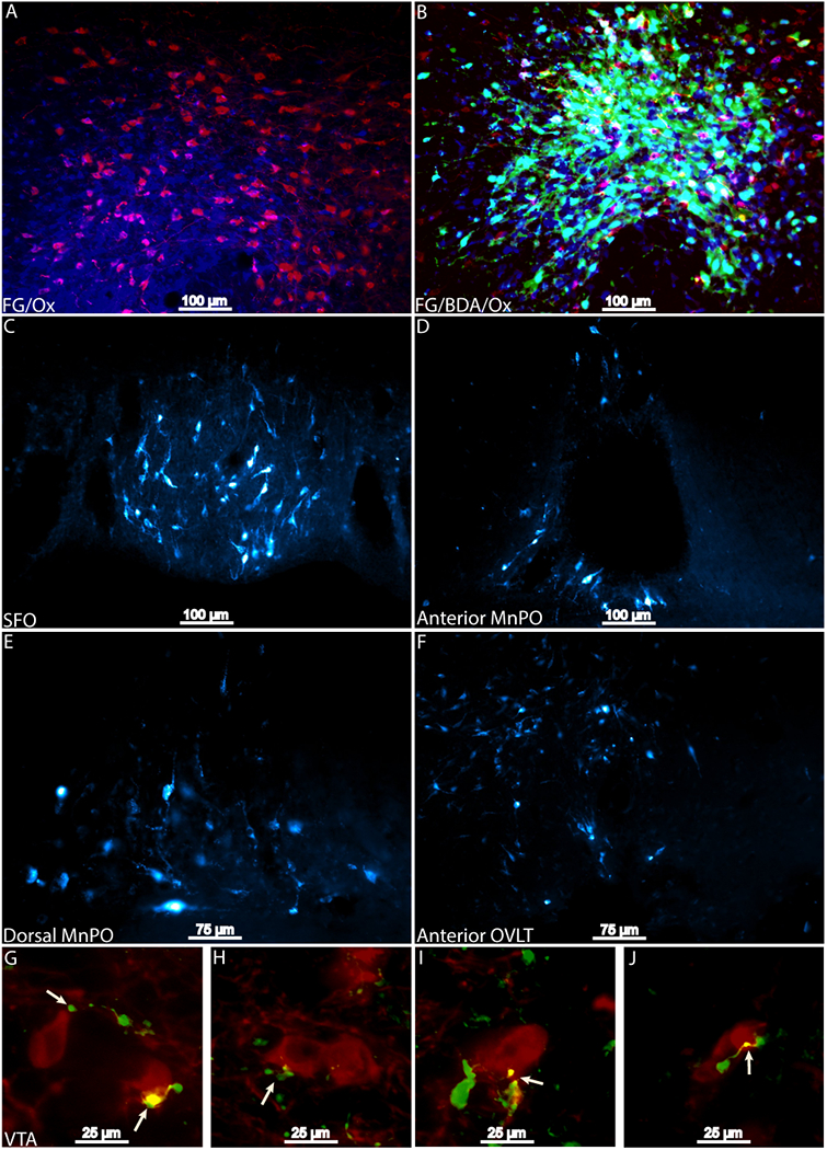
Results from Fluorogold and co-injection of BDA and FG infused into the PeF. Injection sites of 2% FG (blue) into the PeF Ox neurons (red) and a co-injection of 1.33% FG (blue) and 2.5% BDA (green) in PeF Ox neurons (red) are presented in panels A and B, respectively. Retrograde labeling was observed along the entirety of the LT (panels C-F). Axon terminals and varicosities (green) were observed in apposition with tyrosine hydroxylase positive neurons in the VTA (red; panels G-J). Putative synaptic contact is indicated by arrows. BDA, biotinylated dextranamine; FG, Fluorogold; Ox, orexin, PeF, Perifornical Area; SFO, subfornical organ.
