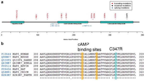
Figure 3.
PRKAR1A gene mutations detected. A. Protein domain structure of PRKAR1A along with mutations observed in the study. Different mutation types are color coded along the protein axis. B. Multiple alignment of PRKAR1A gene sequence from different organisms showing the highly conserved nature of the site of missense mutation C347R. The alignment also shows the close proximity of the missense mutation to the cAMP binding sites. [Color figure can be viewed in the online issue, which is available at wileyonlinelibrary.com.]

