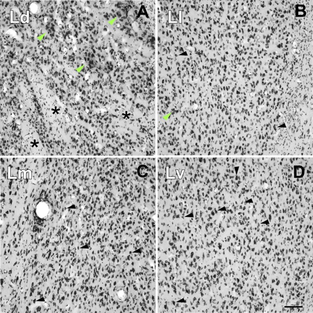
Figure 6.

High-power photomicrographs showing the cytoarchitecture of the lateral nucleus subdivisions. (A) Ld neurons are medium to large sized and clustered (green arrows). Asterisks indicate accumulations of small areas of white matter. (B) Neurons in the Ll have different shapes (black arrows) and are less densely packed than in the Ld (A) and Lm (C), and more densely than in the Lv (D); neuron clusters are rare (green arrow). (C) Neurons in the Lm have a variety of shapes and sizes (black arrows). (D) The Lv is characterized by a relatively low neuronal density with great variability in their sizes and shapes and poorly stained. Scale bar: 100 μm.
