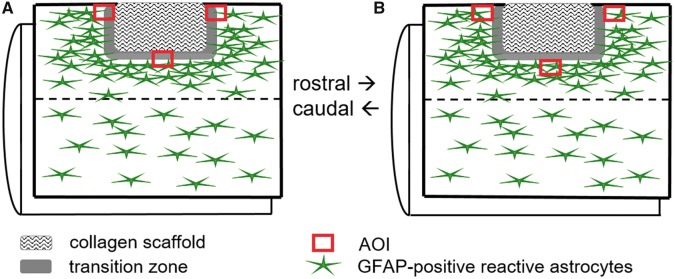
Figure 1.
Schematic diagram illustrating the positions of the AOI for morphometric analyses. A lateral funiculotomy of the right cervical spinal cord followed by implantation of the microstructured type-I collagen scaffold or no implantation (lesion only, not illustrated). (A) Area of interest (AOI) for the quantification of GFAP-immunoreactive astrogliosis defined by the location at the edges of the implanted scaffold. (B) AOI for the quantification of GFAP-immunoreactive astrogliosis defined by the location of the edge of the gliotic tissue

