Figure 6.
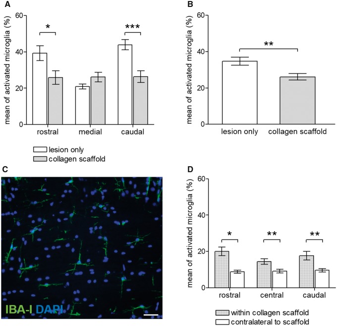
Morphometric analysis of the host microglial/macrophage response to injury and to implantation of the collagen scaffold. (A) Quantification of IBA-I-immunoreactivity demonstrated that the greatest degree of residual microglial staining was at the rostral and caudal lesion–host tissue interfaces. The implantation of collagen scaffold significantly reduced the extent of IBA-I microglial/macrophage staining at both rostral and caudal implant–host tissue interfaces (P < 0.05 and P < 0.001, respectively). (B) Summation of the morphometric analyses at the rostral-, caudal- and medial interfaces demonstrated a significant reduction of the host inflammatory response in response to implantation of the collagen scaffold (P < 0.01). (C) Baseline stain for IBA-I-immunohistochemistry in the contralateral unlesioned spinal cord white matter. Note the small microglial cell bodies with fine, branching processes. (D) Comparisons of the IBA-I-immunoreactivity within the scaffold itself with similarly positioned AOI in the contralateral, unlesioned white matter revealed a statistically significant increase of microglia/macrophage within the scaffold. Values are given as mean±SEM (*P < 0.05; **P < 0.01; ***P < 0.001). Scale bar in (C): 50 μm

