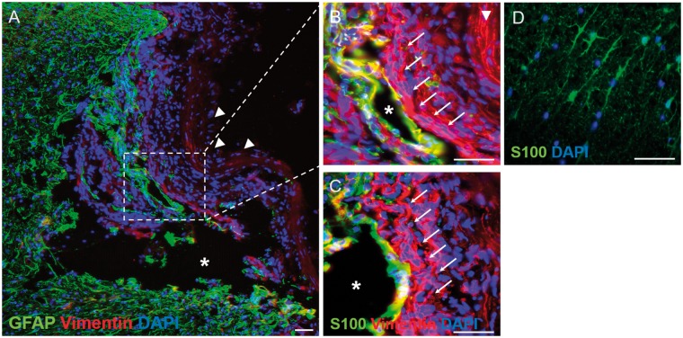
Figure 7.
Immunohistochemical demonstration of the distribution of vimentin, GFAP and S100 within the injury site of the lesion-only group. (A) Low-magnification overview of the injury site stained for vimentin and GFAP. A large cystic cavity (asterisk) can be seen as well as numerous vimentin-positive/GFAP-negative cells. The lesion site appears to have collapsed slightly but the dura mater can still be seen (arrowheads). A protuberance, containing GFAPpositive cells, can be seen penetrating the lesion site, part of which is shown at higher magnification in (B). (B) The GFAP-positive protuberance can be seen surrounding a small cystic cavity (asterisk) and is surrounded by numerous vimentin-positive/GFAP-negative cells that form part of the lateral tissue bridge (arrows), lying next to the inner-most edge of the dura mater (arrowhead). (C) A near adjacent section stained for S100 shows that at this particular level, the vimentin-positive/GFAP-positive cells lining the small cystic cavity were also S100-positive suggesting an astrocytic phenotype, however, the vimentinpositive/GFAP-negative cells of the lateral tissue bridge (arrows) are also S100-negative, suggesting a fibroblast-like phenotype. For a comparison with the baseline staining of contralateral, unlesioned white matter astrocytes, see (D). (D) Baseline stain for astrocytic S100 in a transverse cryosection in the contralateral unlesioned spinal cord white matter. Note the baseline branching morphology of the S100-positive astrocytic profiles. Scale bars (A)–(D): 50 μm

