
Fig 1.
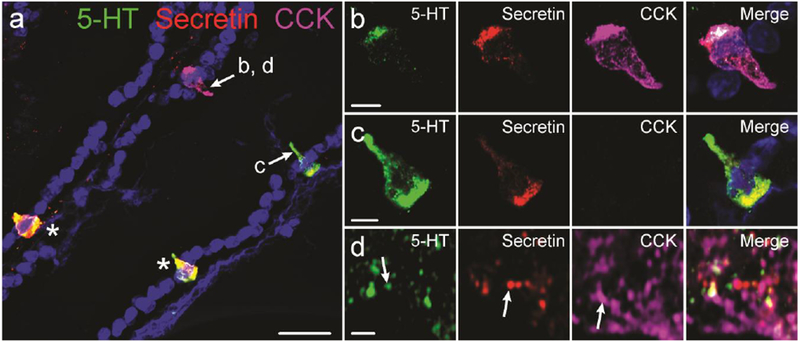
Immunohistochemical colocalization of intestinal hormones. a: 5-HT, secretin, and CCK immunolabelling in mouse duodenum. Cells indicated with arrows are shown in parts b, c, and d. Cells indicated by asterisks are labelled for 5-HT and secretin. Example of a cell containing 5-HT, secretin, and CCK (b), and a cell containing 5-HT and secretin but not CCK (c). Super-resolution image of 5-HT, secretin, and CCK vesicles within an enteroendocrine cell (d). Examples of vesicles in which only one hormone was detected are indicated by arrows. Scale bars: a: 20μm; b, c: 5μm; d: 1μm.