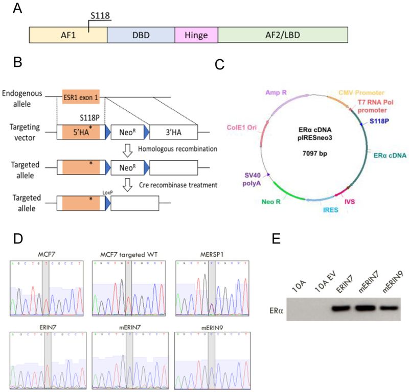
Figure 1: Generation of isogenic cell line models containing ER S118P.
A) Domains of the estrogen receptor-alpha protein and location of the serine 118 residue. AF1=Activation function 1; DBD=DNA-binding domain; AF2=Activation function 2; LBD=Ligand-binding domain. B) Recombinant AAV targeting strategy for knock-in of the variant S118P in ESR1 exon 1 in the ER-expressing cell lines MCF7 and T47D. 5’HA=5’ homology arm; 3’HA=3’ homology arm. C) pIRESneo3 plasmid containing mutated ER cDNA transcript for stable expression of variant ER in the non-ER-expressing cell line MCF10A. D) Sanger sequencing of complementary DNA from the entire panel of cell lines confirms expected heterozygous single base pair change in exon 1 of ESR1. E) Immunoblot analysis of exogenous ER expression for the MCF10A cell panel

