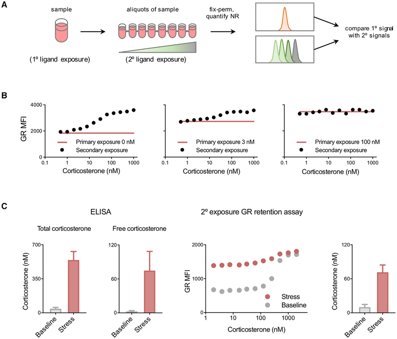
Figure 6. Glucocorticoid Measurement by Ligand Titration Assay.
(A) Protocol for estimation of corticosterone exposure. Cells were cultured in 0, 3, or 100 nM corticosterone (primary exposure), then divided into aliquots and treated with various corticosterone concentrations (secondary exposure). Cells were then perm-fixed, and data were acquired by flow cytometry.
(B) GFP-GR 3617 cells treated as described above. Data are representative of two independent experiments.
(C) Blood samples were collected from mice immediately after disturbance (<2 min, n = 6) or after 15 min of handling stress (n = 5) and divided into aliquots. Plasma was isolated, and total and free corticosterone were quantified by immunoassay (left two panels). Blood was incubated with the indicated corticosterone concentrations, perm-fixed, and stained, and data were acquired by flow cytometry (secondary exposures, right panels). The corticosterone concentrations shown in the rightmost panel were calculated using B cells as described in the STAR Methods. Data are presented as means ± SEM.
See also Figure S3.

