
Fig. 3.
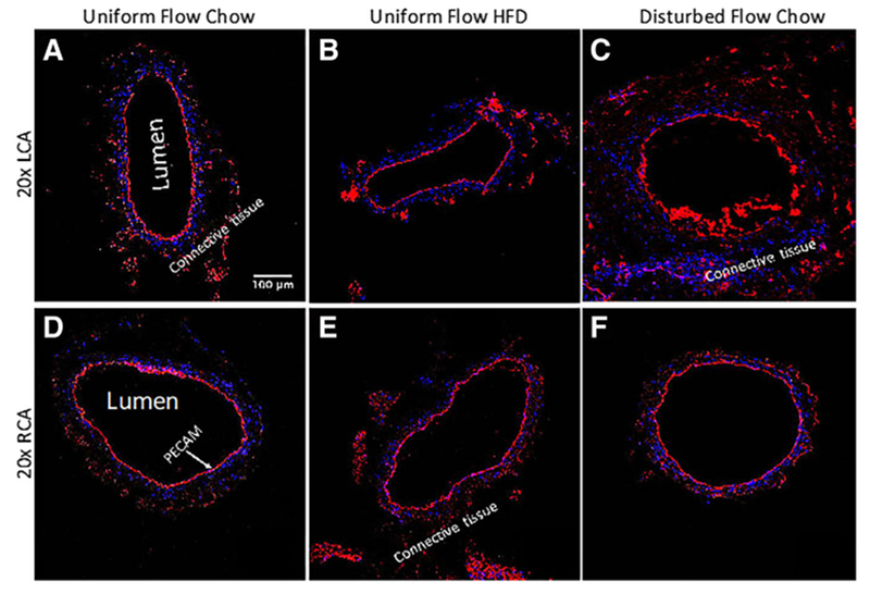
LCA and RCA vessels are immunostained for PECAM (red) to demonstrate the integrity of the endothelial cell layers on the inner vessel walls. Blue signifies DAPI labeled cell nuclei. a 20× image of the LCA obtained from an ApoE-KO mouse with a regular chow diet and exposed to uniform blood flow patterns. b 20× image of the LCA from an ApoE-KO mouse fed an HFD and exposed to uniform flow. c 20× image of the LCA from an ApoE-KO mouse on a regular chow diet and exposed to disturbed blood flow due to partial ligation. d 20× image of the RCA obtained from an ApoE-KO mouse with a regular chow diet. This RCA was exposed to uniform blood flow while its corresponding LCA was also exposed to uniform blood flow. e 20× image of the RCA from an ApoE-KO mouse on an HFD. This RCA was exposed to uniform blood flow while its corresponding LCA was also exposed to uniform blood flow. Finally, f 20× image of the RCA from an ApoE-KO mouse on a regular chow diet. This RCA was exposed to uniform blood flow while its corresponding LCA was exposed to disturbed blood flow