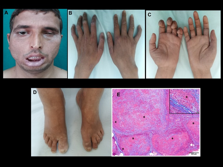
Figure 1.
Case 1: Clinical photographs showing (A) severe bifacial weakness and (B) macular healing lesions over interphalangeal joints. Note the wasting of the interossei muscles; (C) note the asymmetrical wasting and weakness of the hand. Radial cutaneous nerve biopsy; (D) note mild pedal edema and trophic changes of the toes; (E) multiple epithelioid granulomas (*) expanding the nerve fascicle, replacing the contents. Inset shows granuloma with Langhans giant cell (*). Note the fibrosis of the endoneurium. (Masson trichrome, magnification = scale bar). This figure appears in color at www.ajtmh.org.

