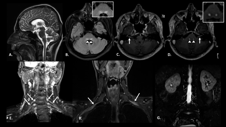
Figure 2.
Case 1: Pretreatment magnetic resonance imaging images: Pretreatment: (A) T2 sagittal section of the brain showing hyperintensity in dorsal pons. (B) Fluid-attenuated inversion recovery (FLAIR) axial section of the brain at the level of facial colliculus shows bilateral symmetrical hyperintensity in the facial nerve nuclei (inset). (C and D) Post-contrast T1 magnetization-prepared rapid gradient-echo axial sections show enhancement of bilateral facial nerves along with their nuclei (inset) in pons. (E and F) Coronal short inversion time inversion recovery (STIR) images reveal thickening of bilateral brachial plexus roots, trunks (E), and divisions (F). (G) Coronal STIR image of the lumbar plexus does not reveal any nerve thickening.

