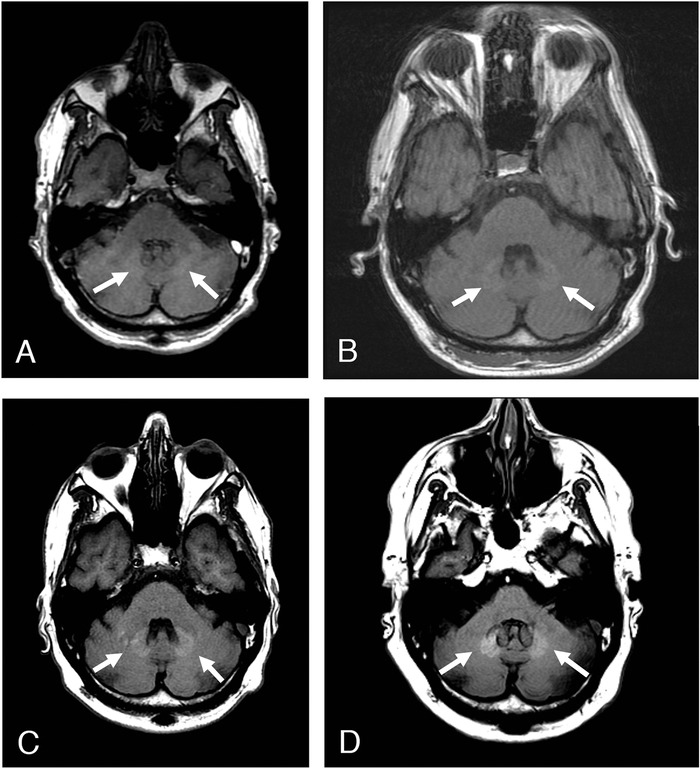
Figure 2.

Sequential axial T1-weighted noncontrast mag-netic resonance images through the posterior fossa of a 52-year-old male with history of multiple sclerosis demonstrate a progressive increase in signal within the dentate nuclei following repeated doses of gadolinium-based contrast agents in (A) December 2011, (B) June 2013, (C) September 2016, and (D) June 2018.
