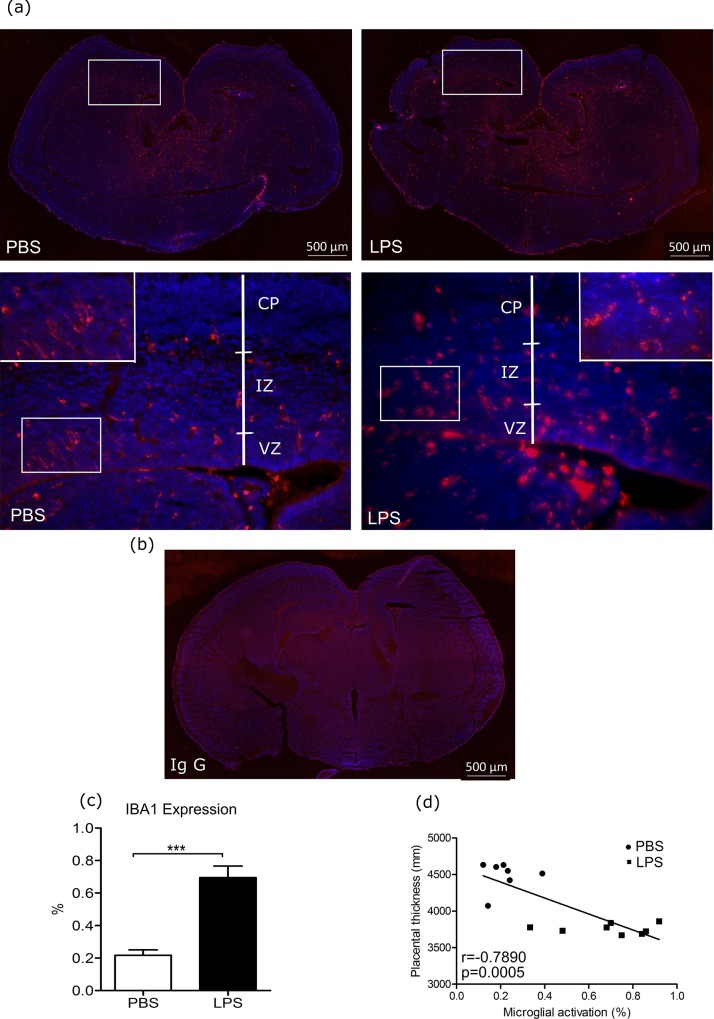
Fig 5. Iba 1 expression in fetal brain and correlation to placental thickness.
(a) Representative images of ionized calcium-binding adaptor molecule 1 (Iba1) expression in fetal brain of E17 mice six hours after lipopolysaccharide (LPS) intra-uterine exposure are presented, the studied areas were the cortical area, including the cortex plate, intermediate zone, and ventricular zone. (b) Negative control image using rabbit Ig G in place of primary antibody. (c) Iba1 positive expression in fetal brains was determined through quantitative analysis as the percentage of Iba1 expression area within the image using Image J (PBS: n = 7, LPS: n = 8). (d) Placental thickness was correlated with microglial activation (Pearson correlation analysis). A Panel: blue represents DAPI stain for nuclei, and red represents Iba1 stain. *** p<0.001; scale bar: 50μm. CP: cortical plate, IZ: intermediate zone, VZ: ventricular zone.

