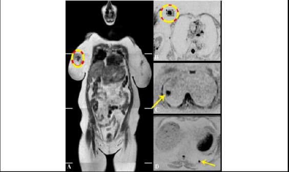
Figure 4-2.

The previous case of metastatic breast cancer; A) WB-MRI coronal T1W image. The primary breast lesion can be seen at upper inner quadrant of the right breast. It is showed by dark signal and irregular outlines; B) Corresponding axial inverted DWI. Breast lesion are shown as a flared out zone of evident diffusion restriction. The mean ADC value of the zone is 0.91 x 10⁻³ mm2/sec; C) & D) Axial inverted DWI at different levels. The pulmonary nodules are shown as rounded focal zones of diffusion restriction at lateral segment of right middle lung lobe C) and posterior segment of left lower lung lobe D) giving a low mean ADC value ranging between 0.97 and 1.03 x 10⁻³ mm2/sec
