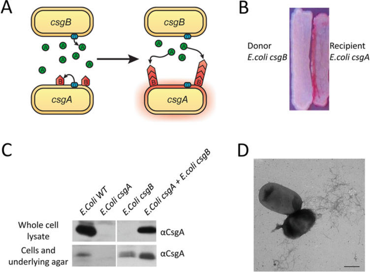
Fig. 2.
Interbacterial complementation between an E. coli csgA mutant and a csgB mutant. (a) A schematic representation of interbacterial complementation. A csgB mutant (the donor) secretes soluble CsgA into the media, which assembles into curli fibers on the cell surface of an adjacent csgA mutant (the recipient) expressing CsgB. (b) A csgA mutant and a csgB mutant were streaked adjacent to each other on a YESCA CR plate and incubated at 26 °C for 48 h. Colonies of the csgA mutant facing the csgB mutant stained red. (c) Western blot analysis was used to detect formation of intercellular curli fibers between an E. coli csgA mutant and a csgB mutant. Overnight cultures of csgA and csgB mutants were mixed in a 1:1 ratio and spotted onto a YESCA plate and a thin YESCA plate. Whole cell lysates and the plugs were collected for western analysis. A wild type E. coli, a csgA and a csgB mutant were used as controls. All the samples were treated with HFIP and probed with anti-CsgA antibody. (d) A mixed culture of E. coli csgA and csgB mutants was spotted onto YESCA plate and grown for 48 h. Curli were detected by EM. Scar bar is 500 nm

