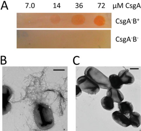
Fig. 5.
Purified CsgA assembles into curli fibers on CsgB expressing cells. (Figure adapted from [46]). (a) CR staining of CsgA–B+ mutant and CsgA–B− mutant overlaid with different concentrations of freshly purified CsgA. Only CsgA–B+ cells overlaid with CsgA protein stained red. (b) Negative-stain EM of CsgA–B+ overlaid with freshly purified CsgA. Fibers were observed on the bacterial surface. Scar bar equals to 500 nm. (c) Negative-stain EM of CsgA–B− overlaid with purified CsgA. No fibers were detected. Scar bar equals to 500 nm

