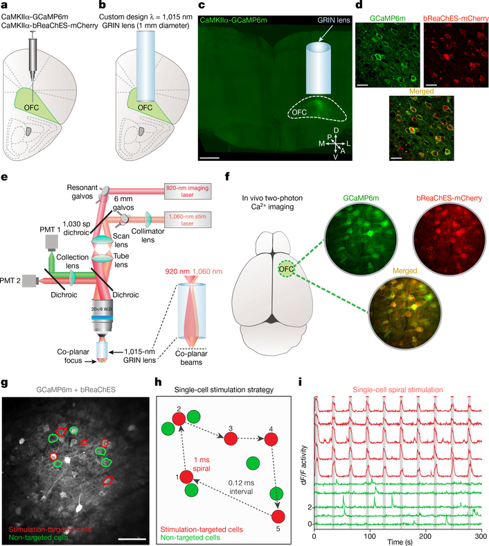
Fig. 1 |. Single-cell two-photon readout and control of OFC neuronal activity in vivo.
a, b, Scheme for targeting OFC with a viral mixture of AAVDJ-CaMKIIα-GCaMP6m and AAV8-CaMKIIα-bReaChES-TS-p2A-mCherry (a) and an implanted GRIN lens (1-mm diameter, 4-mm long, corrected for λ = 1,015 nm) ~2–3 mm ventral (b). Image in a and b is adapted from ref.30. c, Confocal 10× image of a 1-mm thick coronal CLARITY section displaying location of GRIN lens and GCaMP6m expression within the OFC. A, anterior; D, dorsal; L, lateral; M, medial; P, posterior; V, ventral. Scale bar, 500 μm. d, Airyscan 40× images showing co-localization (bottom) of GCaMP6m (top left) and bReaChES-mCherry (top right) in OFC cells. Image from a representative mouse; the experiment was repeated in n = 6 mice with similar results. Scale bars, 20 μm. e, Optical design and dual laser beam paths used for single-cell two-photon resonant scanning (λ = 920 nm; 20–30 mW) and optogenetic manipulations (λ = 1,060 nm; 40–60 mW per spiral target). PMT, gallium arsenide phosphide photomultiplier tube; galvos, galvonomic mirrors; dichroic, dichroic mirror. f, In vivo two-photon visualization of OFC cells co-expressing (bottom) GCaMP6m and bReaChES-mCherry (top). Image from a representative mouse; the experiment was repeated in six mice with similar results. g, Example field of view depicting two-photon stimulation-targeted cells (red circles) and non-targeted neighbouring cells (green circles). Scale bar, 100 μm. h, Diagram illustrating sequential single-cell spiral-stimulation parameters (20-μm spirals, 1-ms spiral duration, 4 revolutions per site, 0.12-ms inter-site interval). i, Ca2+ transients of stimulation-targeted (red) and non-targeted (green) neurons, measured as the relative change in fluorescence (dF/F), demonstrating precise optogenetic activation across multiple stimulations at the single-cell level.

