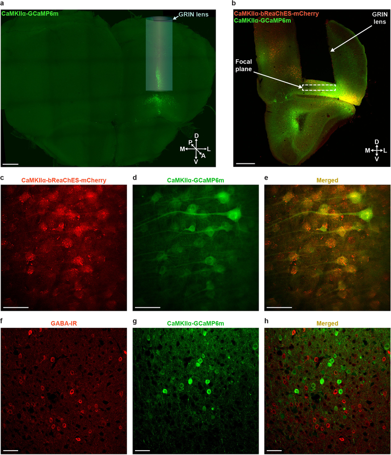
Extended Data Fig. 1 |. Targeting OFC for two-photon cellular-resolution Ca2+ imaging and optogenetic stimulation.
a, Confocal 10× tiled image of a 1-mm-thick coronal CLARITY section displaying the location of the GRIN lens and GCaMP6m expression within OFC. A, anterior; D, dorsal; L, lateral; M, medial; P, posterior; V, ventral. Scale bar, 500 μm. b, Confocal 10× image from a 60-μm coronal slice depicting GRIN lens implantation and viral targeting site within OFC. Scale bar, 500 μm. c–e, Additional representative in vivo two-photon images of OFC cells co-expressing GCaMP6m and bReaChES-mCherry from a different focal plane. Images from a representative mouse; the experiment was repeated in n = 6 mice with similar results. Scale bars, 100 μm. f–h, Confocal 20× images of the OFC showing GABA immunolabelling (f) and CaMKIIα-GCaMP6m expression (g) with minimal overlap (h). Images from a representative mouse; the experiment was repeated in n = 6 mice with similar results. Scale bars, 50 μm.

