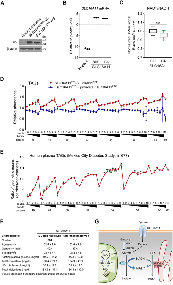
Figure 7. The SLC16A11 diabetes risk haplotype reduces cytosolic NAD+/NADH ratio and increases HUFA synthesis in humans.
(A) Western blot of SLC16A11T2D-V5 and SLC16A11REF-V5 in HEK293 cells, representative gel from one of two independent experiments.
(B) qPCR of SLC16A11 in HEK293 cells expressing empty vector, SLC16A11T2D-V5, or SLC16A11ref-V5.
(C) Cytosolic NAD+/NADH measured with SoNar in HEK293 cells expressing either SLC16A11T2D-V5 or SLC16A11REF-V5.
(D) Intracellular TAGs in HEK293 cells expressing SLC16A11T2D-V5 or SLC16A11REF-V5 (red), or SLC16A11T2D-V5 with or without 1mM pyruvate supplementation (blue). Values are means ± SEM; ***P < 0.001; n = 3 for (B, D), and n = 40 for (C).
(E) Plasma TAGs among carriers of the SLC16A11 risk haplotype (n=364) relative to controls (n=313). Values are means; *P<0.05; **P < 0.01.
(F) Mexico City Diabetes Study clinical characteristics at time of lipid profiling
(G) Schema for interaction between SLC16A11, cytosolic NAD+/NADH, and HUFA synthesis.
See also Figure S7.

