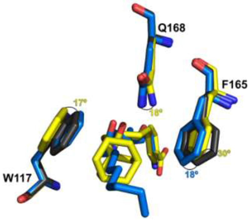
Figure 4. Conformational changes of amino acids caused by the interactions with inhibitors 3 and 5.

Conformational changes for tryptophan117, phenylalanine165 and Glutamine168 for the apo structure (Gray; PDB:5URO) in comparison to the inhibitors 3 (blue) and 5 (yellow). The numbers in blue and yellow show the rotation changes in degrees of the apo structure in comparison with structures in complex with inhibitor 3 and 5, respectively.
