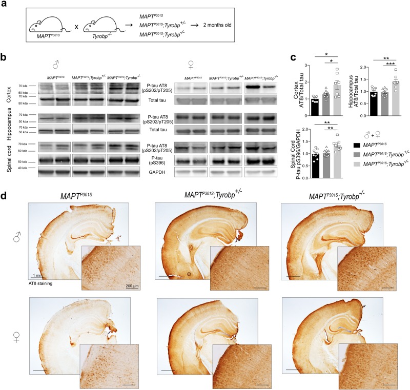
Fig. 1.
TYROBP deficiency rapidly increases phosphorylated tau levels in a MAPTP301S transgenic mouse model of tauopathy. a MAPTP301S mice were crossed with Tyrobp-/- mice, yielding progeny with genotypes of: (1) MAPTP301S mice expressing wild-type TYROBP (abbreviated MAPTP301S); (2) MAPTP301S mice heterozygous for Tyrobp knock-out (abbreviated MAPTP301S;Tyrobp+/-); (3) MAPTP301S mice homozygous for Tyrobp knock-out (abbreviated MAPTP301S;Tyrobp-/-). Male and female mice (2 months of age) were used for this experiment. b Representative western blots of tissues from mice of the indicated genotype were performed using antibodies as indicated: antibody AT8 (detects paired helical filament epitopes of tau phosphorylated at residues serine 202 and/or threonine 205); antibody anti-tau (phospho S396) (detects tau phosphorylated at residue serine 396); antibody T46 (detects total tau); anti-GAPDH. Extracts from cortex, hippocampus and spinal cord from male and female mice of the indicated genotypes were studied. Full-length blots are presented in Supplementary Figure 2. c Densitometric analyses of western blots standardized to total tau or GAPDH. n = 7 (MAPTP301S), n = 11 (MAPTP301S;Tyrobp+/-) or n = 7 (MAPTP301S;Tyrobp-/-) for extracts from cortex; n = 8 (MAPTP301S), n = 9 (MAPTP301S;Tyrobp+/-) or n = 8 (MAPTP301S;Tyrobp-/-) for extracts from hippocampus; n = 8 (MAPTP301S), n = 11 (MAPTP301S;Tyrobp+/-) or n = 7 (MAPTP301S;Tyrobp-/-) for extracts from spinal cord. Material from male and female mice was pooled for analysis. d Representative images of immunohistochemistry with antibody AT8. Hemibrain scale bar = 1 mm; high magnification scale bar = 200 μm. Error bars represent means ± SEM. Statistical analyses were performed using one-way ANOVA followed by Tukey’s post-hoc test, *p < 0.05; **p < 0.01; ***p < 0.001

