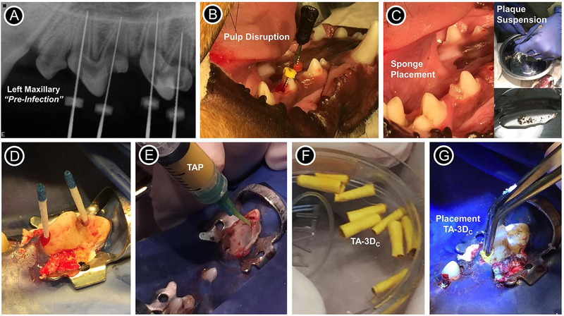
FIGURE 3.
Images showing the periapical lesion induction, disinfection and evoked bleeding procedures performed for the in vivo study. (A) Radiograph from the left maxillary of the dog showing the pre-infection scenario of immature permanent teeth with incomplete root development (i.e., open apices). (B) Pulp disruption performed with a sterile #40 stainless steel endodontic hand file. (C) Placement of sterile sponges inside the pulp chamber and inset images showing the plaque suspension (supragingival plaque) that was mixed with the sponges previously soaked in 0.9% NaCl. (D) Following sponge removal, the root canals were flushed with 10 ml of 1.5% of NaOCl and dried with sterile paper points. (E) For TAP group, a mixture (1 g/ml) containing equal parts of metronidazole, ciprofloxacin, and minocycline pre-mixed with sterile saline was loaded into a plastic syringe and injected into the canals; For tubular 3D triple antibiotic-eluting construct (TA-3DC) group, each TA-3DC was sized to the desired length (F) and press-fitted in the canal spaces using a sterile forceps (G).

