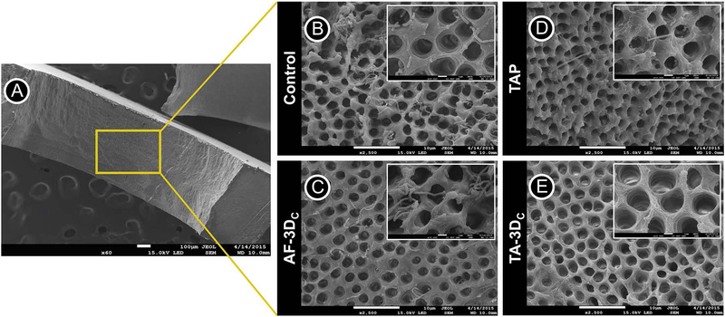
FIGURE 6.
(A) SEM images were obtained from the inner root walls of dentin slices (original magnification ×60). SEM images of A. naeslundii biofilm on the dentin surface (control) (B) treated by tubular 3D antibiotic-free construct (AF-3DC) (C), TAP solution (D), and tubular 3D triple antibiotic-eluting construct (TA-3DC) (E) (original magnification ×2500). Control and AF-3DC group demonstrate viable bacteria on dentin surfaces and inside dentinal tubules. TAP solution and TA-3DC groups eliminated almost all viable bacteria on dentin surface and inside dentinal tubules.

