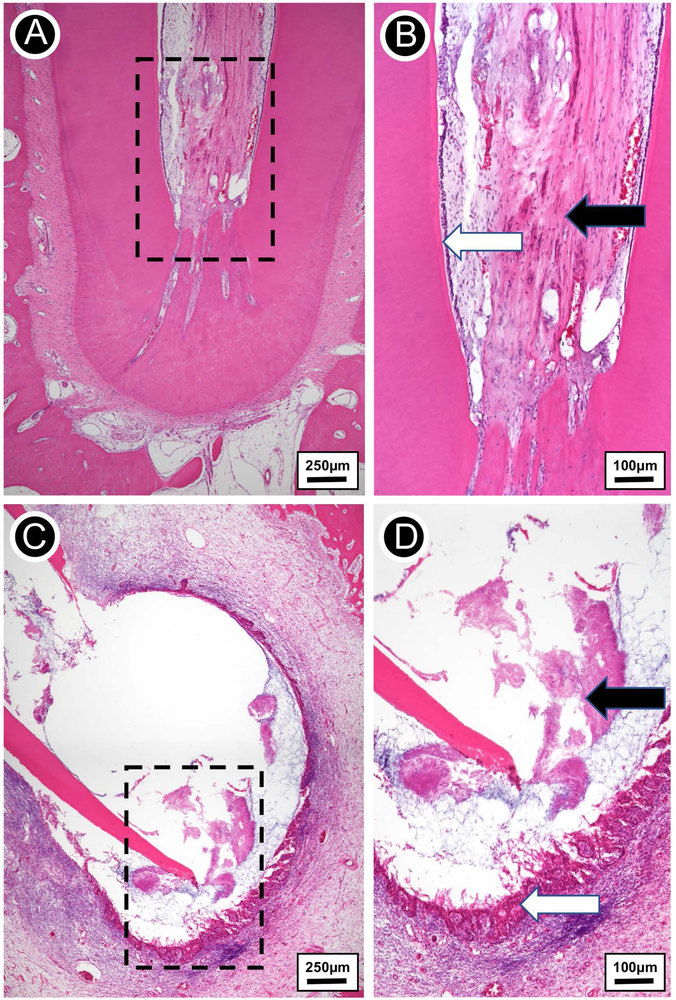
FIGURE 7.
(A) Hematoxylin-eosin stained micrograph of extracted tooth free of infection. (B) Higher magnification view of the rectangular area from A. Vital fibrovascular pulp devoid of inflammation (black arrow), with odontoblasts lining the inner dentin (white arrow). (C) Hematoxylin-eosin stained micrograph of extracted infected tooth showing a periapical lesion comprised of lateral and apical root resorption. (D) A detailed view of the rectangular area from C. Extensive replacement of alveolar bone by chronically inflamed epithelial-lined granulation tissue (black arrow) and filamentous bacterial colonies (white arrow) at the apical third.

