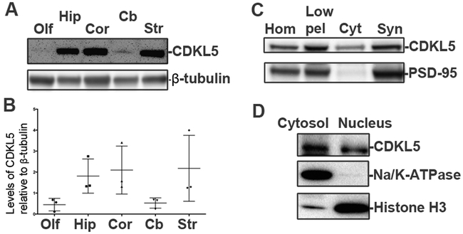
Fig 2: Regional and temporal expression patterns for CDKL5.
A. Western blots for CDKL5 (Sigma) in different regions of the mouse brain as indicated (P28, male, N=3).
B. Levels of CDKL5 relative to β-tubulin in different regions of the mouse brain (P28, male, N=3) (Data ± SEM).
C. Western blot for CDKL5 (Sigma) and PSD-95 (excitatory synaptic marker) in synaptosomal preparations from mouse cortex. (Adult, N=3)
D. Western blot for CDKL5 (Sigma), histone H3 and Na+/K+-ATPase in cytosol and nuclear preparations from primary rat neurons.

