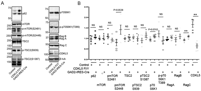
Fig 5: Alterations in levels of components of mTOR signaling pathway with loss of CDKL5 in striatal inhibitory (GAD65-positive) neurons.
A. Western blots for components of the mTOR signaling pathway as indicated in cortical lysates from CDKL5 Fl/Y +/− Gad2-IRES-Cre tissue.
B. Levels of components of the mTOR signaling pathway relative to β-tubulin as indicated in cortical lysates from CDKL5 Fl/Y +/− Gad2-IRES-Cre tissue. (Data presented ± SEM, *=P<0.05, **=P<0.005, ***=P<0.0005, Student’s t-test with Welch correction. N=5 for each genotype. Age 4-20 weeks.)

