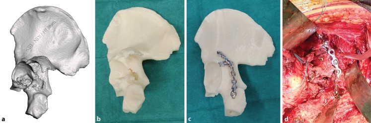
Fig. 1.
An example of a digital three-dimensional (3D) model of a posterior wall acetabular fracture in stereolithography format (a). The 1:1-sized 3D printed model allows the surgeon to accurately appreciate fracture morphology (b). A mirrored model is printed using the opposite intact hemi-pelvis for easy and accurate plate contouring (c). Surgical plan after fracture fixation (d). The implants are placed according to the surgical plan after fracture fixation reduction

