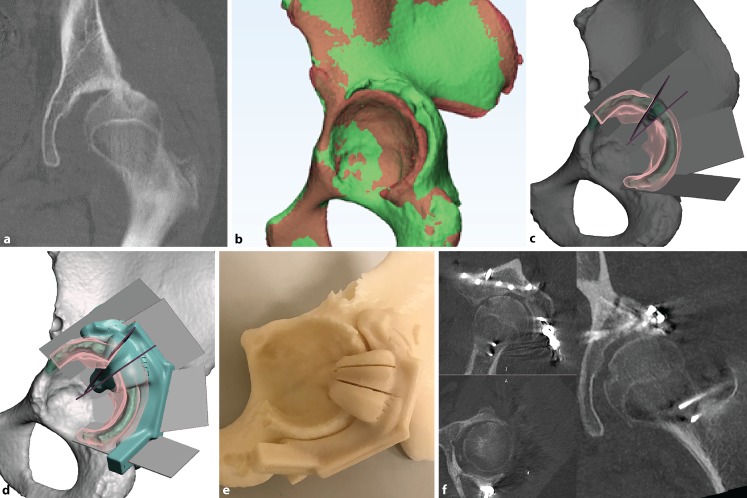
Fig. 4.
A 15-year-old patient with a malunited acetabulum posterior wall with hip subluxation (a). Digital model (green) compared to the mirrored opposite (red) (b). A closing-wedge volume-reducing osteotomy is planned (c) with 3D printed cutting jig (d, e). Postoperative computed tomography showing satisfactory restoration of hip congruency (f)

